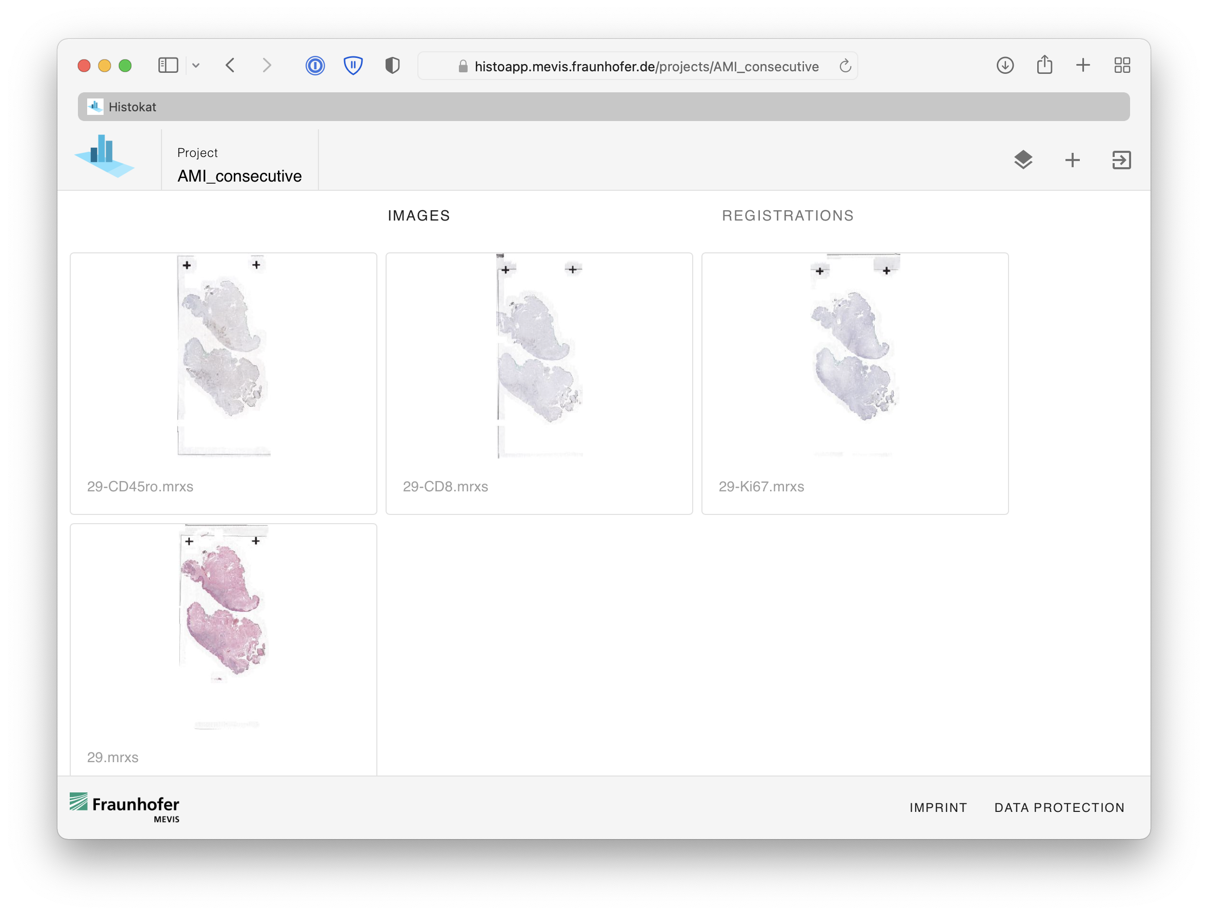
Projects using HistokatFusion
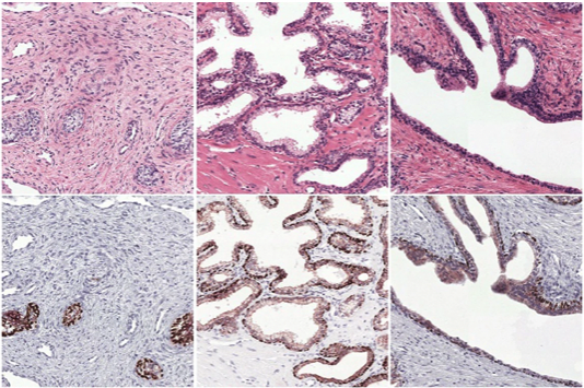
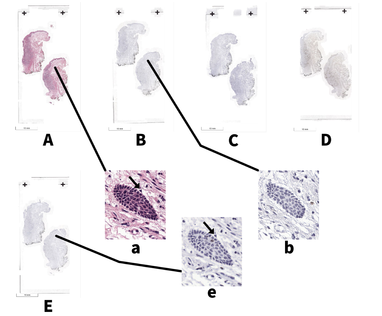
Automatic Annotations using Image Registration
Wouter Bulten et al., Epithelium segmentation using deep learning in H&E-stained prostate specimens with immunohistochemistry as reference standard. Nature Scientific Reports, 2019
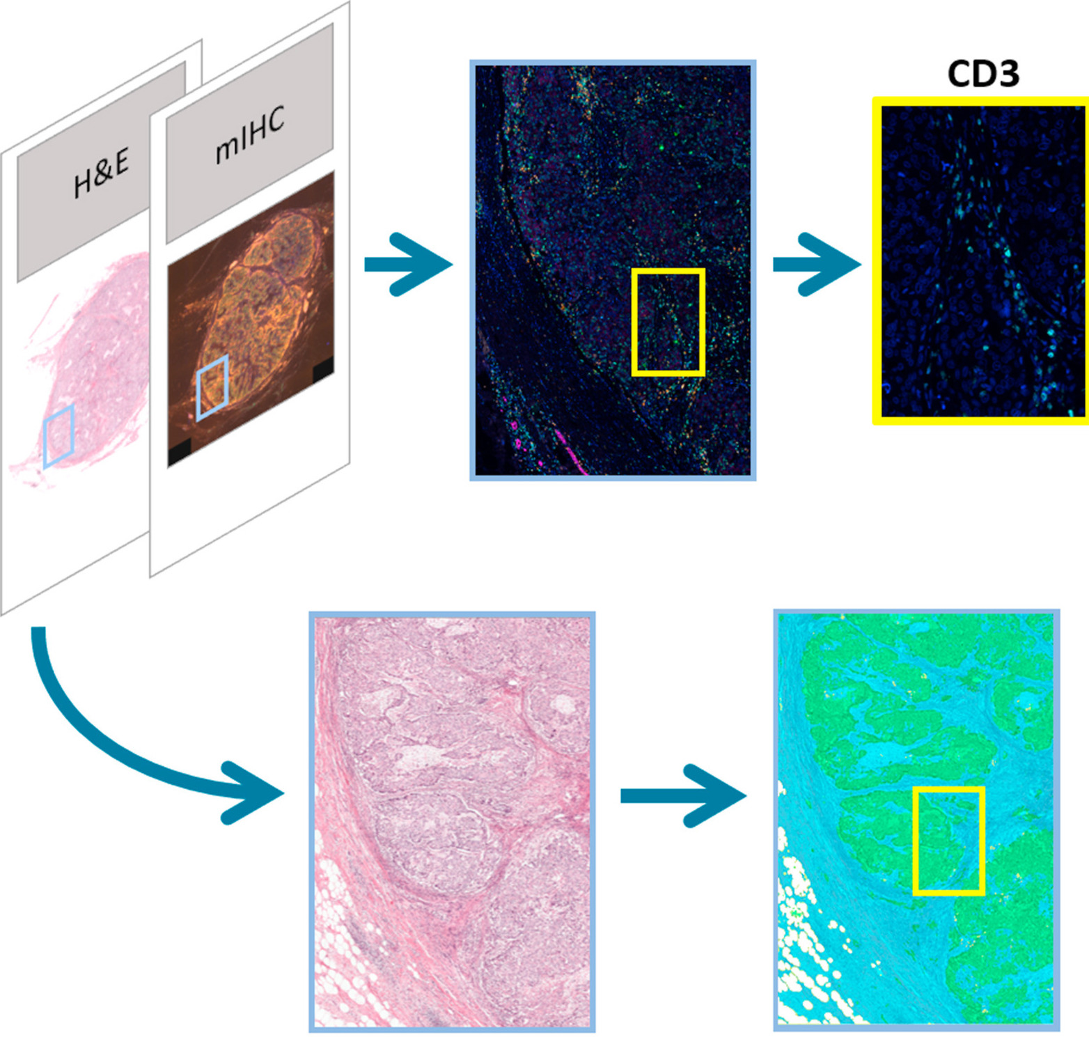
Tumor Inflitrating Lymphocytes in Breast Cancer
Maschenka CA Balkenhol et al., Optimized tumour infiltrating lymphocyte assessment for triple negative breast cancer prognostics, The Breast, 2021
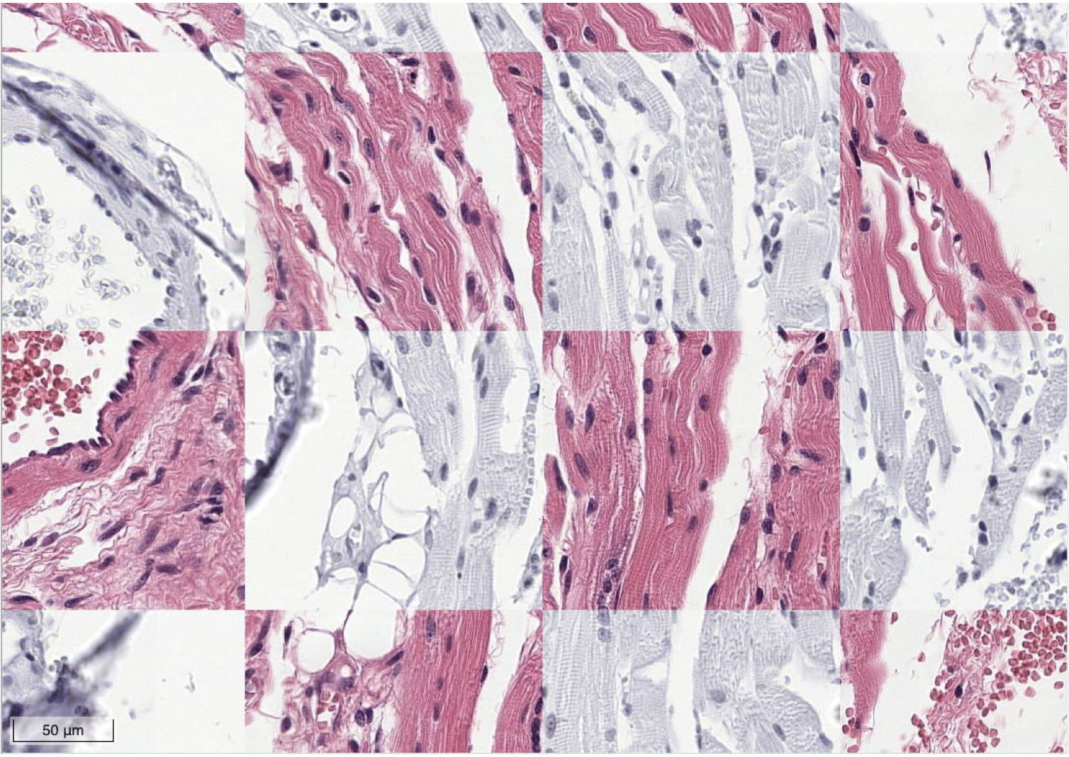
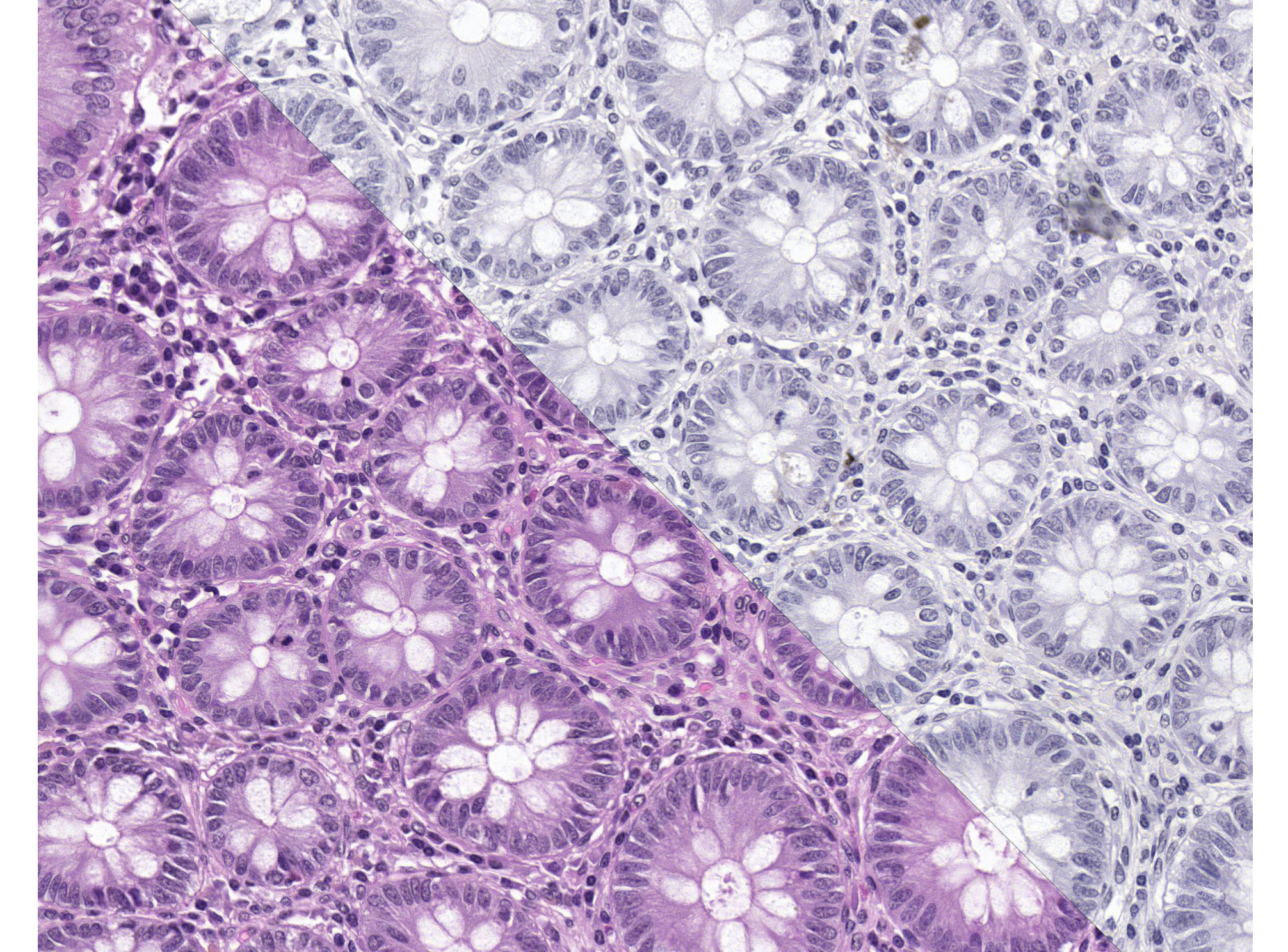
Virtual Staining
C. Mercan et al., Virtual Staining for Mitosis Detection in Breast Histopathology, IEEE 17th International Symposium on Biomedical Imaging (ISBI), 2020